Our principles in varicose vein care
We apply the latest methods to treat varicose vein disease. Every treatment is preceded by advanced duplex ultrasound diagnostics. After surgery we schedule regular follow-up visits and use additional injection therapy to remove any remaining veins.
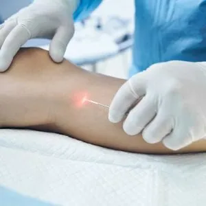
Laser vein surgery
Fewer complications, less time in bandages and a more aesthetic result compared with classic stripping.
Our guide walks you through laser vein surgery from preparation and sedation to aftercare – no incisions (only a puncture), with faster recovery and better long-term results than foam alone.
Patient guide and benefits
This information is for patients preparing for laser vein surgery. At A+B Clinic we have performed vein operations since 1991 and used laser since 2007. Avoiding a groin incision means fewer complications, less post-op discomfort, faster recovery and a shorter compression period than classic surgery; compared with foam injections, long-term results are better with fewer punctures and less inflammation.
Assessment before surgery
Personal consultation and duplex ultrasound determine the best approach. We issue lab and routine test requests; please share results by phone or in person, email them and, if possible, bring a printout.
Pre-op preparation
Surgery slots are typically Wednesday and Friday mornings with individual arrival times. Do not eat beforehand; if the start is later, have a light snack no later than six hours before and stop drinking two hours before, while hydrating well earlier (no alcohol). Take your usual medicines as agreed and bring them with you; keep the skin on the leg dry and pack sleepwear and slippers for the few hours on site.
Surgery day and sedation
Reception welcomes you, you complete the anaesthesia questionnaire during the short wait, a nurse shows your room, we mark the target veins with ultrasound, give a blood-thinning tablet, then you change into paper underwear. An experienced anaesthesiologist provides twilight sedation (not spinal, not deep anaesthesia): we place an IV in the left arm, give fluids and sedation, and monitor with ECG, blood pressure cuff and pulse oximeter; you fall asleep before the procedure starts.
How the procedure runs
Under ultrasound we insert a laser fibre through a tiny puncture to seal the main trunk with heat. Curvy side branches are removed with injection sclerotherapy, and deeper perforators are closed with adhesive. After preparation, one leg takes about 30–60 minutes, both legs 50–80 minutes; at the end, while already awake, we wheel you back to the bed – most patients do not even recall this.
After surgery
We give an infusion and pain relief if needed. You sleep off the sedative but wake easily; about 2–3 hours after finishing you are fully awake, then after a check you stand up, get dressed and go home accompanied. The same day do not drive, sign documents or make important decisions because of the sedatives.
Control visits
The first control is scheduled on the surgery day; the next day or within 2–3 days we change the bandage and check the area with ultrasound. Do not unwrap the bandage before then; you may shower using a plastic cover. Afterwards we change the wrap and scan weekly.
Compression and movement
From day 7 you may remove the bandage and stocking for washing; wash the operated leg with soap, avoid soaking, then after drying rewrap firmly with padding and pull on the stocking (insert video here). From day 14 you may remove it overnight and wrap again in the morning; most wear it for about four weeks, sometimes shorter or longer, occasionally only on part of the limb. At first do not load the leg—stand and move while seated; from week 3 take longer walks, and after a month light sport helps the treated veins seal well.
Need help?
For any concern or unexpected event call Dr. Imre Bihari (+36 30 9605 855) or Dr. Péter Bihari (+36 30 399 4529).
- Incision-free laser vein closure with less discomfort and a shorter bandage period
- Comprehensive assessment, anaesthesiologist-led sedation and ultrasound-guided laser fibre
- Can be combined with injection side-branch treatment and perforator care for durable results
- Ambulatory care (few hours in clinic)
- Call Dr. Imre Bihari – <a href="tel:+36309605855">+36 30 9605 855</a>
- Call Dr. Péter Bihari – <a href="tel:+36303994529">+36 30 399 4529</a>
FAQ
Most patients resume light work after one to two days of rest while wearing a compression stocking.
Online booking currently unavailable.
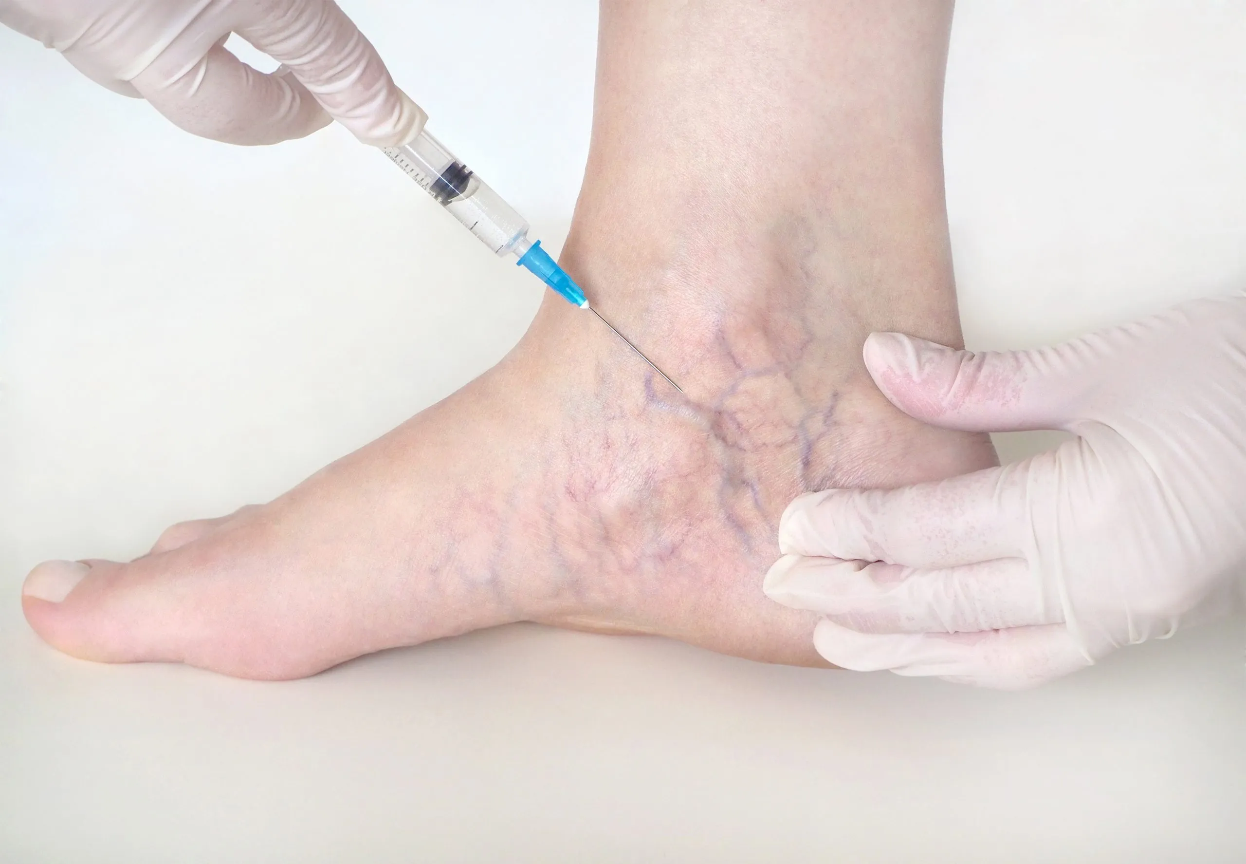
Injection vein therapy (sclerotherapy)
Very nice aesthetic result.
Effective from the finest spider veins to larger varices by sealing the diseased vein with a specialised solution or foam; performed as an outpatient treatment.
How sclerotherapy works
We inject a solution that targets the inner lining of the vessel so the walls adhere and the vein is absorbed. Using foam allows us to treat larger trunks as well, making the method effective from spider veins to broader varices.
Evidence and setting
Sclerotherapy has more than half a century of clinical experience behind it. With today’s proven agents the results are reliable worldwide. Sessions take place as outpatient visits that last little longer than a consultation, so you can stay in your normal work and family routine.
Comfort and risks
Minor inconveniences such as compression bandages or stockings, mild inflammation or temporary pigmentation may appear, yet serious complications are extremely rare. Varicose veins can recur, but repeat sessions and careful follow-up keep the cosmetic result long-lasting.
When we use foam or liquid
For larger veins we use ultrasound guidance and decide individually whether to inject liquid or foam. This helps the treated area heal quickly while you can continue everyday movement immediately.
- Ultrasound-guided foam therapy for larger veins
- Quick treatment with minimal discomfort
- Results can be maintained with repeat sessions
FAQ
Compression bandages or stockings are recommended for the first weeks after treatment to keep the vein walls sealed.
Online booking currently unavailable.
Spider vein treatment
Visible cosmetic improvement through a tailored series of gentle treatments.
Injection-based sclerotherapy that restores an even skin tone for spider veins, telangiectasia and small reticular veins.
Why spider veins respond well
Spider veins – whether fine telangiectasia or reticular veins – respond exceptionally well to sclerotherapy. The carefully dosed solution seals the targeted vessels so they gradually fade and the complexion regains a youthful appearance.
Gentle technique under guidance
We work with ultra-fine needles under ultrasound guidance to reach even the smallest branches. Each session may involve dozens of tiny injections that patients tolerate well; after the visit we recommend short-term compression bandaging or stockings while you resume light everyday activity.
Maintaining the cosmetic result
Because spider veins can recur, we plan maintenance sessions when necessary and discuss realistic expectations, possible side effects such as transient pigmentation and when to combine the treatment with laser or foam techniques for the most even result.
- Cosmetic results continue to refine over the following months
- Treats even thread-like vessels with minimal downtime
- Maintenance or combined laser therapy keeps the outcome stable
Online booking currently unavailable.
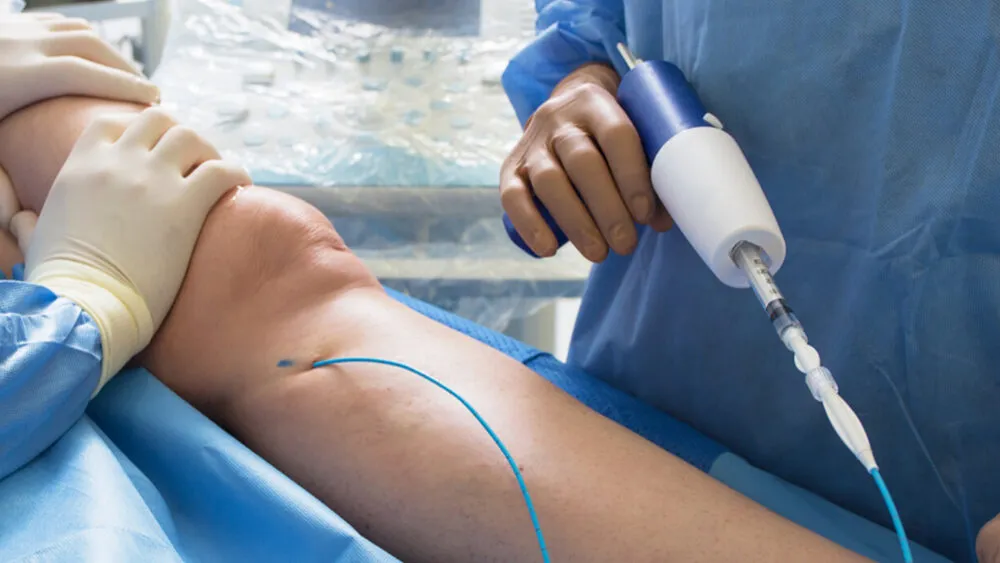
Adhesive vein closure
Single puncture, minimal discomfort, quick return to daily life.
The simplest known vein operation: no pain, no general or spinal anaesthesia, and no need for compression bandages or stockings.
What is this method?
The simplest vein operation we know: no pain, no anaesthesia beyond a tiny local numbing of the puncture, and no stocking or bandage afterwards – almost exactly what most vein patients dream of. We work on an operating table in sterile conditions; studies across Europe and the US show good results.
How does it proceed?
Through a single puncture we advance a catheter into the vein, guide it with ultrasound, then in several steps deliver small amounts of special adhesive. The one puncture receives mini anaesthesia and needs nothing more: no major local anaesthetic, no spinal needle, no general anaesthesia. You feel nothing of the glue delivery; you may feel gentle pressure when we compress the vein walls, but it is not painful. The puncture is tiny, needs no stitches, the procedure takes about 15 minutes and after a few minutes of rest you continue daily activities. Fewer unpleasant sensations and fewer potential complications – surgeons have used this adhesive safely for 50 years, and treating both legs in one session is often advantageous.
Drawbacks
Relatively few cases qualify: very large or very small veins are unsuitable, and even on one leg about 20% of veins may remain for later injections. Europe and the US have tested it widely with good outcomes, but follow-up so far covers only a few years. Mild, short-lived inflammation occurs in about 10% of cases. The specialised material costs more than other newer methods, yet many patients in Hungary have already chosen it for its benefits.
When is it ideal?
We mainly recommend it when visible varicose change is mild but open source veins could worsen quickly; closing them can make the smaller, barely dilated veins retract or disappear. It is useful to slow deterioration, for elderly or co-morbid patients with complicated veins, and if you want a rapid, painless fix while postponing treatment of any residual veins. It can be justified for manual workers, before holidays or travel, and for overweight patients to avoid the discomfort of bandages or tight stockings. If you want to avoid anaesthesia and significant pain relief, an ultrasound consultation clarifies your options.
- Single puncture with mini anaesthesia, about a 15-minute procedure
- No general or spinal anaesthesia, no bandage or stocking
- Adhesive proven safe for 50 years, rare mild inflammation
- Ultrasound selects target segments; residual veins can be injected later
- Ideal if you want a quick, painless solution or wish to avoid compression
FAQ
Mild irritation can appear in about 10% of cases and usually subsides within a few days at the injection site.
Online booking currently unavailable.
Targeted perforator vein care
Our internationally recognised work confirmed that sealing perforator veins with adhesive is safe and effective.
Managing incompetent perforator veins with adhesive or mini-surgical techniques to prevent recurrence of varicose veins.
Targeted care for perforator veins
Perforator veins link the superficial system under the skin with the deeper veins beneath the muscle. These short veins pierce the muscle at a right angle rather than running parallel to the skin. Normally blood flows from the superficial veins through the perforators into the deep system. If a perforator is insufficient, the flow reverses: when the muscles contract, blood is pushed from the deep veins back into the superficial ones, stretching them and causing varicosities. In many cases insufficient perforators drive recurrence or even leg ulcers despite otherwise successful trunk treatment, so whenever the pre-op ultrasound shows an insufficient perforator, we address it during surgery.
How we treat perforators
Previously we treated perforator veins surgically through a small (2–3 cm) incision to locate and tie them off. Today we prefer a less invasive technique: under ultrasound guidance we puncture the target vein with a needle and inject a small amount (0.1–0.2 ml) of adhesive that seals the vessel and eliminates its harmful effect. Because closure requires only one puncture, it can be performed as an ambulatory procedure with minimal discomfort and very good outcomes.
- Focus on Dodd and pelvic perforator pathology
- Ambulatory care with rapid wound healing
- Can be combined with laser or injection therapy
Online booking currently unavailable.
Leg ulcer management
A leg ulcer always has a cause that must be treated to achieve lasting healing.
Not just a leg wound: we identify the underlying condition, treat the ulcer and prevent recurrence.
What counts as a leg ulcer?
A leg ulcer is a chronic wound on the lower leg that appears without injury. Patients sometimes link the start to a tiny trauma, yet a disproportionately large wound forms or healing stalls, typically in the lower third of the leg, most often just above the inner ankle.
Causes and underlying diseases
Many conditions can cause a leg ulcer: venous or arterial circulation issues, lymph drainage problems, diabetes, dermatological diseases or combinations of these. Treatment is therefore often complex, requiring collaboration among a vascular surgeon, dermatologist, internist, endocrinologist, wound-care specialist and lymph therapist.
The role of varicose disease
A common cause is dilated superficial veins with faulty valves. Blood fails to travel upward to the heart and instead flows downward toward the ankle; deep veins try to drain it, but used blood continually refluxes, the skin becomes firmer, darkens, inflames and eventually breaks down.
Benefit of surgery
In such cases removing the superficial veins by classic or laser surgery stops used venous blood from flowing back into the leg. Circulation improves, wound healing speeds up and the chance of recurrence falls.
- Find the root cause across venous, arterial, lymphatic or metabolic factors
- Wound care with modern dressings plus compression and lymph support
- Vascular, internal medicine and dermatology teamwork for lasting healing
Online booking currently unavailable.
Lymph therapy and oedema care
Gentle, stroking lymph massage opens new lymph pathways to reduce swelling without causing pain.
Manual and mechanical lymphatic drainage with compression bandages and stockings to achieve lasting reduction of swelling.
When lymph therapy helps
Disorders of the lymphatic system frequently cause ankle and leg swelling; without treatment the oedema can harden and progress to elephantiasis.
Manual and mechanical drainage
We combine manual lymph drainage with mechanical compression devices to gently mobilise the trapped fluid, then use bandages or stockings to maintain the reduction.
Exercises and skin care
During the intensive phase we apply bespoke bandaging, teach targeted exercises and skin care so the swelling decreases day by day.
Maintenance to prevent relapse
A personalised maintenance plan with home routines, regular reviews and booster sessions keeps the oedema under control for the long term.
- Early detection of lymphatic circulation disorders
- Tailored compression protocols
- Long-term home self-care guidance
Online booking currently unavailable.